





Bone Grafting
Precision-Guided, Patient-Centered Care
Personalized Planning and Clinical Care
Data Collection and Assessment
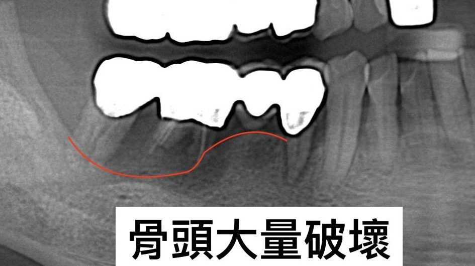
Cone-beam CT and intraoral scans may be used to assess the teeth and bone and create a 3D model. The dentist uses these records to evaluate the area requiring augmentation, plan the procedure, and select an appropriate surgical approach.
Surgical Procedure
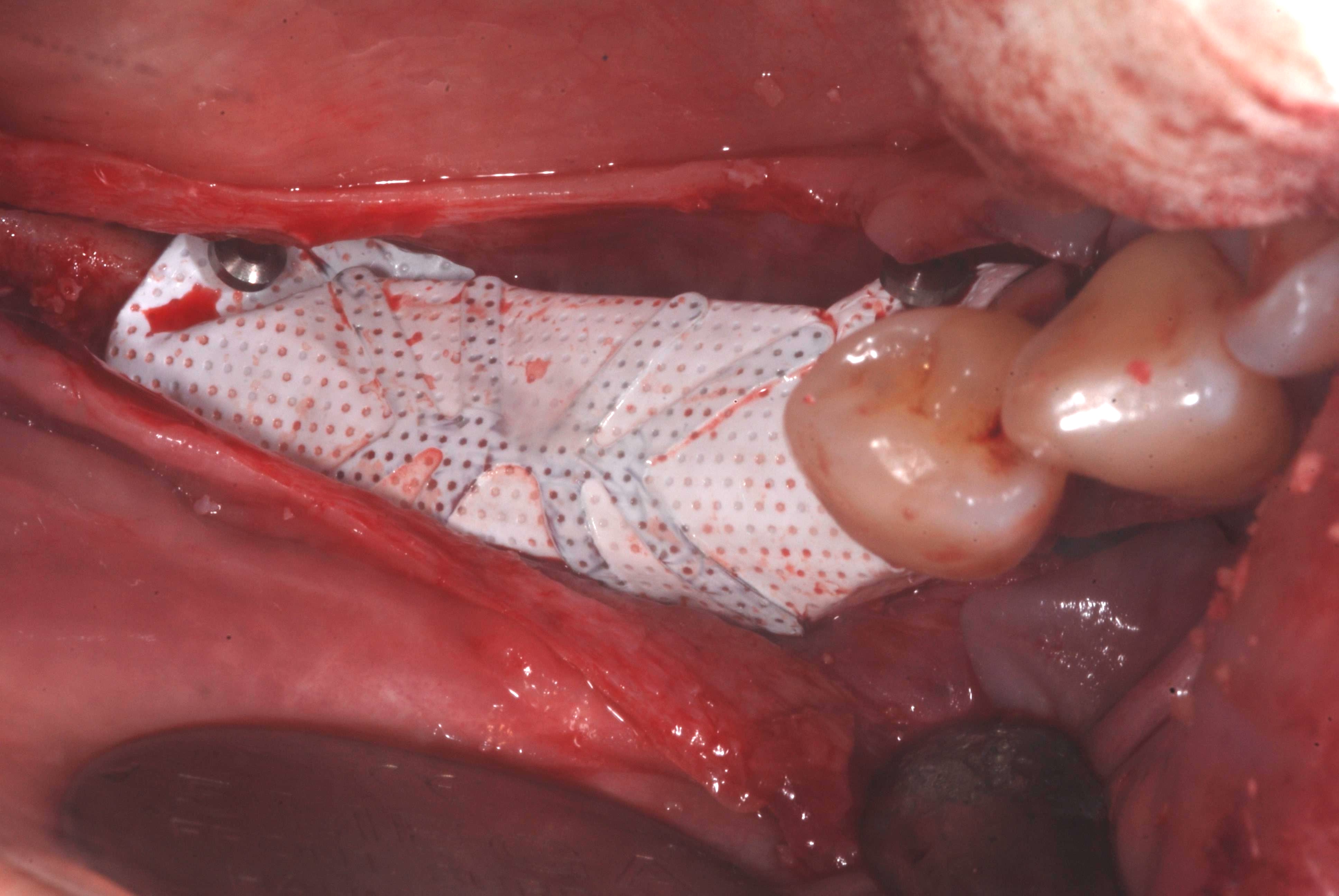
The treating dentist reconstructs the deficient area with grafting material. Depending on the clinical condition, a titanium-reinforced framework or membrane may be used to maintain space and support guided bone and periodontal tissue regeneration.
Completing Treatment
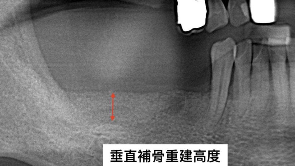
A follow-up visit is usually scheduled about one week after surgery for suture removal and wound evaluation. Further treatment is planned after healing and clinical stability are confirmed.
Navident Dynamic Navigation Implant Surgery (Minimally Invasive da Vinci Robotic Arm Implant Surgery) FAQs
Q8. Who is suitable for minimally invasive implant surgery? Can it be performed if there is insufficient bone?
Most patients who want to restore chewing function after tooth loss, trauma, or extraction due to periodontal disease are suitable for an assessment for da Vinci robotic arm minimally invasive implant surgery. Before surgery, however, the dentist must use a 3D CT scan to carefully assess whether the height, width, and density of the jawbone are sufficient to support the artificial tooth root. If bone is severely insufficient, bone grafting may be needed first, followed by implant placement once the bone has healed and stabilized.
Q11. Can an implant be placed immediately after a tooth is extracted, or must I wait for the bone to heal?
Some cases with favorable conditions, such as intact bone at the extraction site and no obvious inflammation or infection, may allow immediate implant placement at the same time as extraction. With its small incisions and limited effect on surrounding tissue, a minimally invasive technique is more favorable for this type of procedure. However, if bone is insufficient or inflammation or infection is present, it is still recommended to wait for the bone and soft tissue to heal and stabilize before reassessing the appropriate timing for implant placement.
Q12. What examinations are needed before implant surgery? Is a 3D CT scan necessary?
With 3D CT images, the dentist can precisely measure jawbone height, width, and bone density, while clearly identifying the exact locations of nerves and blood vessels. This information is an indispensable basis for implant navigation planning and directly affects the safety and accuracy of surgery.